Our last blog on endotoxins is about the importance of endotoxins in general. Here, we introduce the structure of one of the best investigated endotoxins – lipopolysaccharide (LPS).
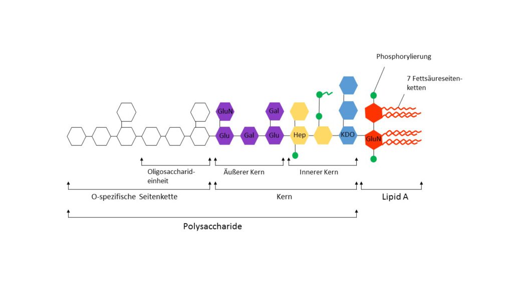
LPS occurs only in the cell wall of Gram-negative not Gram-positive bacteria, respectively. It contributes to the structural integrity of the bacteria and provides protection against environmental conditions like salts, lipophilic antibiotics and many more.1 Molecularly, LPS consists of the hydrophobic, highly conserved lipid A, the hydrophilic core polysaccharide components (core) and the highly variable O-specific polysaccharide side chain (Figure 1). In most cases, the O-responsible side chain is made up of repetitive oligosaccharide units that lead to strain specific structural diversity. The different strain specific O serotype is determined by the organization and interconnection of the oligosaccharide units. In this way the O serotype is the main criterion for the serotyping classification of Salmonella. Evolutionarily, the various O-Serotypes of the Enterobacteriaceae have developed to escape the host’s immune system by varying the surface structure of the bacteria. By displaying LPS on the bacterial surface it acts as an antigen and is also known as O-antigen.2

Figure 1. General structure of LPS. The highly conserved Lipid A consists of 7 fatty acid side chains, 2 GluN = N-acetylglucosamine residues and 2 modifications (usually phosphorylation, this can vary in a strain and growth dependent manner). Moreover, the fatty acid chains can vary slightly in length and composition (mostly capronic, lauric, myristic, palmitic or stearic acid). The core area (core) consists of 1-4 KDO, GluN, Hep = heptose (most often L-glycero-α-D-manno-heptopyranose), Glu = glucose and Gal = galactose and, depending on the species, the number of sugars and modification (most often pyrophosphate or 2-aminoethyl phosphate) vary slightly. In most wild types, the inner core, in particular the KDO = 2-keto-3-deoxy-octonate area, is also relatively well preserved. The O-specific side chain is relatively variable and consists of species and strain-specific repetitive oligosaccharide units (hexoses: rhamnose, galactose, glucose, mannose and one or more dideoxy sugars such as abequose, colitose, paratose or tyvelose), whereby the individual sugars can be partially modified. Adapted from Silverman and Ostro, 1998.
It is an immunological classification that specifies which antibody recognizes which strain. Different strains can have common antigens and can be recognized by the same antibody. A serotype important for testing is the Escherichia coli serotype O113: H10: K–, whose endotoxin was used for the development of the international endotoxin standard.3
A wide range of LPS molecules of different lengths occur in wild-type strains, an effect that is based on the step-by-step bio-synthesis of the polysaccharide side chain.4 It is also possible to generate strains with mutations in LPS synthesis in the laboratory, which leads to different LPS depending on the mutation variants that do not occur in nature. Ra, Rb, Rc, Rd and Re are names for polysaccharide chains of different lengths (Figure 2). Ra and Re are the two most extreme variants, where Ra denotes the longest polysaccharide side chain (without O-specific part), while Re variants consist only of lipid A and 3 units of KDO.5 Lipid A is obtained from Re strains by hydrolysis of LPS, since hydrolysis becomes more complex with increasing polysaccharide side chain length. The diphosphorylated (toxic) or the monophosphorylated (non-toxic) variant of lipid A can be obtained by hydrolysis of the side chain.6–8 If the fatty acid chains of lipid A are removed, this also leads to a loss of toxicity.9

Figure 2. LPS mutants and variability. The closer a structure is to Lipid A, the higher its degree of conservation (or the lower the variability between different strains). Rough strains are mutants produced in the laboratory and do not occur in nature. The term smooth, or rough, comes from the corresponding colony morphology when growing on a suitable solid nutrient medium. Ra variants are the longest while Re variants have the shortest LPS. Adapted from Silverman and Ostro, 1998.
Literatur
1. Bacterial endotoxic lipopolysaccharides. (CRC Press, 1992).
2. Methods in microbiology: D. W. Ribbons. Vol. 18: […]. (Academic Press, 1985).
3. 10-178.pdf. at <http://www.nibsc.org/documents/ifu/10-178.pdf>
5. Raetz, C. R. Biochemistry of endotoxins. Annu. Rev. Biochem. 59, 129–170 (1990).